
Australiformis
| Australiformis | |
|---|---|
| Scientific classification | |
| Domain: | Eukaryota |
| Kingdom: | Animalia |
| Phylum: | Acanthocephala |
| Class: | Archiacanthocephala |
| Order: | Moniliformida |
| Family: | Moniliformidae |
| Genus: | Australiformis Schmidt and Edmonds, 1989[1] |
| Species: | A. semoni
|
| Binomial name | |
| Australiformis semoni | |
| Synonyms | |
| |
Australiformis is a monotypic genus of acanthocephalans (thorny-headed or spiny-headed parasitic worms) containing a single species, Australiformis semoni, that infests marsupials in Australia and New Guinea. Its body consists of a proboscis armed with hooks which it uses to pierce and hold the gut wall of its host, and a long trunk. This genus resembles species in the genus Moniliformis but is characterized by a lack of spiral muscles in the outer wall of the proboscis receptacle. The proboscis is armed with 12 rows of 13 to 15 hooks which are used to attach themselves to the small or large intestines of the host. The female worms range from 95 to 197 millimetres (3.7 to 7.8 in) long, virtually all of which is the trunk, and 1.75 to 3.5 millimetres (0.069 to 0.138 in) wide. There is pronounced sexual dimorphism in this species as females are around twice the size of the males whose trunks range from 46 to 80 millimetres (1.8 to 3.1 in) long and 2 millimetres (0.079 in) wide. Infestation by A. semoni may cause debilitating inflammation of the stomach (gastritis) with granulomatous ulcers.
Taxonomy[edit]
The taxonomic history of A. semoni is complex. It was originally named Echinorhynchus semoni by Linstow in 1898,[2] and then moved to Gigantorhynchus by Porta in 1908[3] and Johnston in 1909, later moved to Prosthenorchis by Travassos in 1917,[4] then renamed Moniliformis semoni by Johnston and Edmonds in 1952[5] before taking the present name and genus by Schmidt and Edmonds in 1989.[6][1] The genus is monotypic, the only species, Australiformis semoni (Linstow, 1898)[a], being necessarily the type species.[7] Linstow named the species semoni after the German zoologist who discovered it, Richard Semon.[2]
The morphological traits of a simple, double-walled proboscis receptacle, eight cement glands (which are used to temporarily close the posterior end of the female after copulation) each with a giant nucleus, the brain at the posterior end of proboscis receptacle, and dorsal and ventral lacunar canals place this genus confidently in the order Moniliformida. The genus Australiformis Schmidt and Edmonds, 1989 was created for Moniliformis semoni as this species differed from other species in Moniliformis and the other genera of the family Moniliformidae, Promoniliformis, because it lacked spiral muscles in the outer wall of the proboscis receptacle. The parasitizing of marsupials is also a unique trait of this genus among Acanthocephala.[1] No genetic testing has been conducted on this species to confirm this classification.[1]
| Archiacanthocephala |
| Phylogenetic reconstruction for select species in the class Archiacanthocephala based on a 28S rRNA gene comparison from Gomes et. al (2019) and a 18S rDNA gene comparison from Amin et al. (2020).[8][9] Unlike the other species shown, no genetic testing has been conducted on Australiformis semoni to confirm this classification which is inferred based on morphological traits.[1] |
Description[edit]
| Measurements[1] | Female (mm) | Male (mm) |
|---|---|---|
| Length of proboscis | 0.640–0.800 | 0.600–0.840 |
| Width of proboscis | 0.280–0.320 | 0.200–0.288 |
| Length of proboscis receptacle | 1.2–1.58 | 1.0–1.7 |
| Width of proboscis receptacle | 0.3–0.48 | 0.32–0.36 |
| Length of neck | 0.225–0.240 | 0.150–0.240 |
| Width of neck at base | 0.200–0.270 | 0.200–0.270 |
| Length of trunk | 95–197 | 46–80 |
| Width of trunk | 1.75–3.5 | 2 |
| Length of lemnisci | 25–27 | 20–25 |
| Size of anterior testis | 2.7–3.2 x 0.75–1.0 | |
| Size of posterior testis | 2.5–3.2 x 0.75–1.0 | |
| Size of cement glands | 0.64–1.2 x 0.44–0.72 | |
| Size of Saefftigen's pouch | 1.28 x 0.48–0.64 | |
| Size of eggs | 0.080–0.086 x 0.035–0.044 | |
| Distance from the uterine bell[b] to genital pore | 1.95–2.2 |
A. semoni consists of a proboscis covered in hooks, a proboscis receptacle, and a long trunk. There is pronounced sexual dimorphism in this species; the females are around twice as long as the males (up to approximately 20 centimetres (7.9 in) in females and 8 centimetres (3.1 in) in males). The proboscis is long and swollen at the anterior end and tapers rapidly to a narrow base. The proboscis is armed with 12 rows of 13 to 15 hooks. The first three to four hooks in each row are large, aligned in straight rows, and have bifid roots whereas the other 10 to 12 posterior hooks are small rootless spines arranged in spirals down the proboscis. The first hook of each row is 40 to 56 micrometres (0.0016 to 0.0022 in) long, the second is 50 to 60 micrometres (0.0020 to 0.0024 in) long, the third is 42 to 50 micrometres (0.0017 to 0.0020 in) long, the fourth is 42 to 54 micrometres (0.0017 to 0.0021 in) long and the remaining spines are 30 to 60 micrometres (0.0012 to 0.0024 in) long. At the base of the proboscis is a double-walled proboscis receptacle with a smooth outer wall, lacking spirally arranged muscle fibers, and a large space between the walls. The brain is located near the posterior end of the proboscis receptacle with retinacula (a band of thickened deep fascia around tendons that holds them in place) piercing the proboscis receptacle wall laterally. Proboscis retractor muscles pierce the posterior end of the proboscis receptacle.[1]
The trunk is not pseudosegmented, is very thin at the anterior end and thickest at the posterior end averaging only a few millimeters in width. The main longitudinal lacunar canals are dorsal and ventral, with the dorsal canal being conspicuous and the ventral canal being very narrow. The transverse commissural canals are evenly spaced and connect to the main longitudinal canals. The lemnisci (bundles of sensory nerve fibers) are long, slender, twisted, and coiled in the body cavity (not attached distally to the body wall), and contain 10 to 15 giant nuclei each. They extend between one quarter to one third the length of the body.[1]
The eggs are oval with three apparent membranes. The outer membrane is thick with the exception of the anterior end where it is thin. The outer membrane is often indented and the posterior end is usually covered in small dots on the outer surface with a knob on the inner surface. The second membrane is very thin and the third membrane is thick. The males have a sensory pore on each side of the neck. Males have eight oval cement glands, each with a single giant nucleus, and possess a Saefftigen's pouch just behind the testes.[1] The testes are oval in tandem and found near the posterior end of the trunk. The genital pore is located at the terminal end of the trunk in both sexes.[1]
Distribution[edit]
The distribution of A. semoni is determined by that of its hosts. A. semoni has been found in several states of Australia, including Queensland, New South Wales, and Tasmania. This parasite has also been found in Boroko, Papua New Guinea. The type locality is the Upper Burnett River region in south-eastern Queensland.[1]
Hosts[edit]

The life cycle of an acanthocephalan consists of three stages beginning when an infective acanthor (development of an egg) is released from the intestines of the definitive host and then ingested by an arthropod, the intermediate host. Although the intermediate hosts of Australiformis are not known, without exception for the order Moniliformida, this intermediate host is an insect. When the acanthor molts, the second stage called the acanthella begins. This stage involves penetrating the wall of the mesenteron or the intestine of the intermediate host and growing. The final stage is the infective cystacanth which is the larval or juvenile state of an Acanthocephalan, differing from the adult only in size and stage of sexual development. The cystacanths within the intermediate hosts are consumed by the definitive host, usually attaching to the walls of the intestines, and as adults they reproduce sexually in the intestines. The acanthor are passed in the feces of the definitive host and the cycle repeats. There are no known paratenic hosts (hosts where parasites infest but do not undergo larval development or sexual reproduction) for Australiformis.[12]
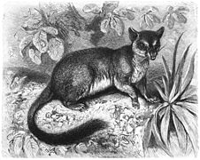
A. semoni parasitizes Australian and New Guinean marsupials including the type host, southern brown bandicoot (Isoodon obesulus) and related species such as the northern brown bandicoot (Isoodon macrourus), long-nosed bandicoot (Perameles nasuta), striped bandicoot (Perameles gunnii), common echymipera (Echymipera kalubu), and brush-tailed phascogale (Phascogale tapoatafa). A. semoni infests these hosts by using hooks on their proboscis to pierce and hold the wall of the small and large intestines.[1] A. semoni has also been found with the anterior end embedded in the mucosa of the stomach in the striped bandicoot. This infestation, which all observed cases contained 5 or fewer individual worms, may cause debilitating ulcerative granulomatous gastritis, a form of gastritis (inflammation of the stomach) characterised by ulcers and granuloma (an aggregation of macrophages that forms in response to chronic inflammation).[13] Juvenile worms were found in the accidental host (an organism that generally does not allow transmission to the definitive host) brown antechinus (Antechinus stuartii).[1] There are no reported cases of A. semoni infesting humans in the English language medical literature.[11]
- Hosts for Australiformis semoni
-
 The Southern brown bandicoot is the type host of A. semoni.
 -

-

-
_2,_Vic,_jjron,_09.01.2013.jpg)


Notes[edit]
- ^ A binomial authority in parentheses indicates that the species was originally described in a genus other than Australiformis.
- ^ a funnel like opening continuous with the uterus.
- ^ There are no known aberrant human infections for A. semoni species.[11]
References[edit]
- ^ a b c d e f g h i j k l m n Schmidt, G.D.; Edmonds, S.J. (1989). "Australiformis semoni (Linstow, 1898) n. gen., n. comb. (Acanthocephala: Moniliformidae) from marsupials of Australia and New Guinea". The Journal of Parasitology. 75 (2): 215–7. doi:10.2307/3282769. JSTOR 3282769. PMID 2926590.
- ^ a b von Linstow, O.F.B. (1898). "Nemathelminthen von Herrn Richard Semon in Australien gesammelt". Denkschriften der Medizinisch-Naturwissenschaftlichen Gesellschaft zu Jena (in German). 8: 471–472. Retrieved 23 March 2020.
- ^ Porta, A. (1908). "Gli acantocefali dei mammiferi. Noto preventiva". Archives de parasitologie. 12 (2): 268–282.
- ^ Travassos, L. (1917). "Contribuigoes para o conhecimento da fauna helmintolojica brazileira. VI. Revisao dos acantocefalos brazileiros. Parte l. Fam. Gigantorhynchidae Hamann, 1892". Memórias do Instituto Oswaldo Cruz (in Portuguese). 9: 5–62. doi:10.1590/S0074-02761917000100001.
- ^ Johnston, T.H.; Edmonds, S.J. (1952). "Australian Acanthocephala No. 9". Transactions of the Royal Society of South Australia. 75: 16–21. Archived from the original on 21 March 2023. Retrieved 21 March 2023.
- ^ "Moniliformida Schmidt, 1972". Integrated Taxonomic Information System (ITIS). 23 November 2019. Archived from the original on 3 May 2023. Retrieved 30 January 2020.
- ^ Amin, O.M. (19 September 2013). "Classification of the Acanthocephala". Folia Parasitologica. 60 (4): 273–305. doi:10.14411/fp.2013.031. PMID 24261131.
- ^ Nascimento Gomes, A. P.; Cesário, C. S.; Olifiers, N.; de Cassia Bianchi, R.; Maldonado, A.; Vilela, R. do V. (December 2019). "New morphological and genetic data of Gigantorhynchus echinodiscus (Diesing, 1851) (Acanthocephala: Archiacanthocephala) in the giant anteater Myrmecophaga tridactyla Linnaeus, 1758 (Pilosa: Myrmecophagidae)". International Journal for Parasitology: Parasites and Wildlife. 10: 281–288. doi:10.1016/j.ijppaw.2019.09.008. PMC 6906829. PMID 31867208.
- ^ Amin, O.M.; Sharifdini, M.; Heckmann, R.A.; Zarean, M. (2020). "New perspectives on Nephridiacanthus major (Acanthocephala: Oligacanthorhynchidae) collected from hedgehogs in Iran". Journal of Helminthology. 94: e133. doi:10.1017/S0022149X20000073. PMID 32114988. S2CID 211725160.
- ^ CDC’s Division of Parasitic Diseases and Malaria (11 April 2019). "Acanthocephaliasis". www.cdc.gov. Center for Disease Control. Archived from the original on 8 June 2023. Retrieved 17 July 2023.
- ^ a b Mathison, BA; et al. (2021). "Human Acanthocephaliasis: a Thorn in the Side of Parasite Diagnostics". J Clin Microbiol. 59 (11): e02691-20. doi:10.1128/JCM.02691-20. PMC 8525584. PMID 34076470.
- ^ Schmidt, G.D. (1985). "Development and life cycles". In Crompton, D.W.T.; Nickol, B.B. (eds.). Biology of the Acanthocephala (PDF). Cambridge: Cambridge Univ. Press. pp. 273–305. Archived (PDF) from the original on 22 July 2023. Retrieved 16 July 2023.
- ^ Lenhaus, C.; Obendorf, D.; Wright, F.H. (1990). "Veterinary aspects of Perameles gunnii biology with special reference to species conservation". In Clark, T.W.; Seebeck, J.H. (eds.). Management and conservation of small populations. Brookfield, Illinois: Chicago Zoological Society. pp. 89–108. ISBN 0-913934-16-X. Retrieved 23 March 2020.